
![]()
IV. Was Prototaxites a lichen? |
I. The
fossil II. A fungus? (Hueber) III. An alga? (Schweitzer) V. Miscellaneous VI. Literature and credits |
In 2002 Marc-AndrÚ Selosse from Paris published a reaction on Hueber's paper on Prototaxites. He valued Hueber's research very much, but he also had serious doubts about his conclusions. Particularly on the following three points.
1. The remains of the reproductive structures, described by Hueber, are incomplete and not convincing. Furthermore it is peculiar that no spores have been found, not even in the direct vicinity of embedded specimens of Prototaxites.
2. The huge size of the fruiting body of the fungus cannot be clearly explained. Organisms become big in competition with other organisms, but plants in the Early Devonian didn't grow much higher than 50 cm. Besides it is doubtful how the giant fungus could find enough food. A rule of thumb is that the biomass in forests at a certain level of the food chain is at most about 10% of the next lower level. The fungi have a mass of about 10 % of the layer of humus in which they live. Prototaxites, however, seems to have had about the same mass as the layer on which it lived.
3. Prototaxites became extinct in the Late Devonian. Hueber thinks that this was possibly caused by predation by animals and by competition of trees and bushes. The latter, however, doesn't make sense, for fungi and plants are not food competitors and a fungus is not dependent on light. Plants only bring more food for the fungi.
Selosse thinks it possible to overcome these objections by assuming that Prototaxites was a lichen. That is a cohabitation of a fungus and an alga or a cyanobacteria with mutual benefit (symbiosis). Fungi can gain minerals and water from the soil with their mycelium, whereas algae and cyanobacteria can produce nutrients from carbon dioxide and water by assimilation. Furthermore the alga/cyanobacteria and the fungus can give each other protection in very difficult circumstances. The alga/cyanobacteria is sometimes called photobiont. Selosse thinks that the fotobiont in Prototaxites was an alga.
According to Selosse, the photobiont must have been situated near
the outside of the youngest growth increment, because there light could
be caught. The alga in the older (inside) parts died for lack of light, but
the tubes stayed intact and because of their firm structure they gave
stiffness to the trunk. The so-called skeletal hyphae must have been the tubes
of the alga in Selosse's view.
Nowadays algae which form tubes without septa do exist. In them growth is
concentrated in the top and to prevent the flowing back of the protoplasm
a temporary septum is formed. The rest of the tube is empty. It is possible
that the alga belonged to this type. Moreover, green algae can produce chemicals
which strengthen the walls.
Retallack & Lanting published a paper in 2014 in which they, describing a trunk
with branches and remains of the original surface, show that Prototaxites must have been a lichen. The
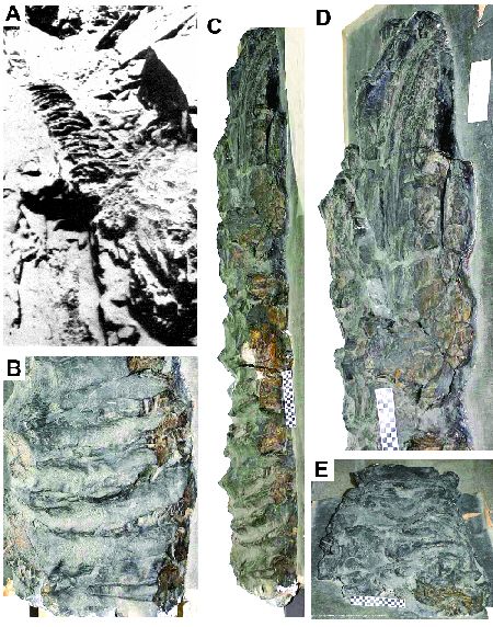
Schunnemunk Tree: Prototaxites loganii.
A. Photo of the branched top at the time of the excavation in 1898; B.
The wrinkeled trunk near the top. C. Upper part of the trunk. D. Detail
of the bases of the branches. E. Separate basal piece of a branch
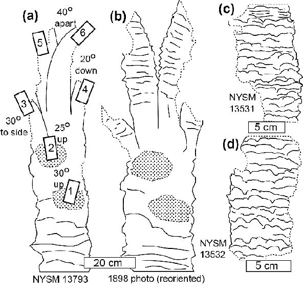
with wrinkles. Sketches
of the fossil. A. The trunk as it lies in the museum. B. The trunk as
it was photographed in 1898. C and D. Fossil branches found in
2011. Reprinted with
permission from Mycologia. ®The Mycological Society of America
The fossil they used, is a complete trunk of Prototaxites
loganii
from the Middle Devonian (approx. 386 million year old), that was
found in a small quarry in the State of New York at the end of the 19th
century (see the figures below). The trunk is in the New
York State Museum in Albany under the name of Schunnemunk Tree. It's
length is 8.83 m and in the upper part it has six branches which are
each about 1 m long and 9 cm thick.
The
trunk is silicified and at transverse sections it shows the same
image of thick and thin tubes (hyphae) as other specimens of Prototaxites.
The basal parts of the branches have been well preserved. Still in 2011
coalified remains of three branches were recovered. At the trunk and at the branches there are irregular, coarse wrinkles.
The outermost layer
At some places at the base of the trunk the coalified outside has been preserved rather well.
Under the whole surface there proved to be a narrow gap of about
25 Ám, which was only interrupted at small invaginations (see the
pictures below). From the transverse sections and the SEMs it turned
out that nests with small spheres ("coccoid cells") were situated
under the invaginations. The coccoids are visible in clusters of strongly
branched thin hyphae. The diameter of such a sphere is about 6 Ám. Some
of them are whole, others are penetrated by hyphae. The researchers
think that these coccoid cells are the remains of the photobiont.
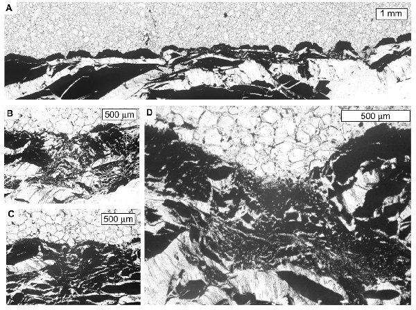 Transverse sections of the coalified outer layer of the trunk. A. Ribs and invaginations of the surface and underneath an elongated cavity, sometimes interrupted at invaginations. B, C and D. Nests of hyphae with in them badly preserved little spheres. These can be seen more clearly below. |
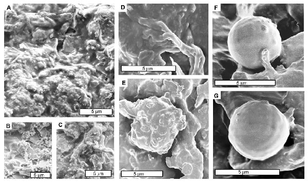 Scanning
electron micrographs (SEMs) of clusters of coccoid cells in the middle of and
sometimes penetrated by hyphae. These coccoid cells must be the fossil
photobiont. Reprinted with permission from Mycologia. ®The Mycological Society of America. |
As the
alga or cyanobacteria needed light, it was necessarily living at the
outside of the trunk. The thick and thin hyphae are part of the fungal
component. At the concentric rings spots with branching hyphae have
been described bij Hueber (2001), but no coccoid cells have been found
in these spots. Retallack & Lanting think these are old nests of
the photobiont from which the alga/cyanobacteria has disappeared.
Furthermore a kind
of radially directed structures have been found, running from those
'old nests' to the outer side. Retallack & Lanting speculate that
the photobiont has migrated via these routes to the outer side. The
figure below gives an impression of their idea of the structure of a
trunk of Prototaxites.
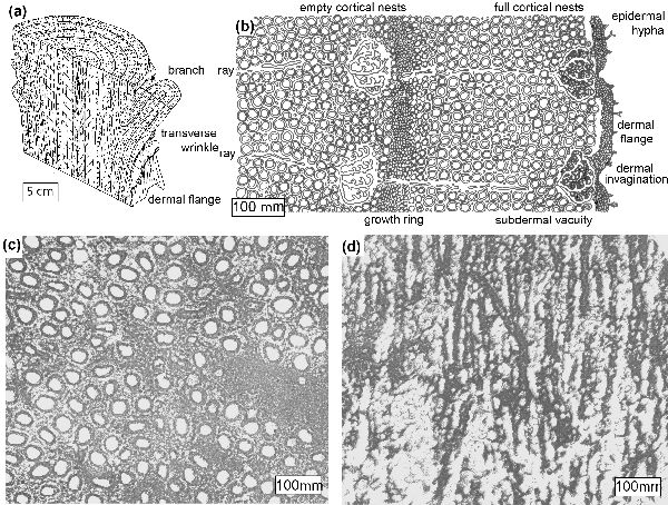 A.
Sketch of the structure of a trunk with branch and wrinkles. B.
Schematic drawing of the transverse section of the outermost layers of
the trunk. C. Transverse section of the silicified trunk with hypae. D.
Longitudinal section with hyphae. Reprinted with permission from Mycologia. ®The Mycological Society of America |
 Prototaxites according to Retallack & Landing. |
Considering
the diameter of the coccoid cells Retallack & Landing think that
the phobiont must have been an alga. The diameter of cyanobacteria in recent
lichens is commonly larger than 10 Ám, whereas the diameter of algae
happens to be lesser than 10 Ám. The coarse wrinkles at the trunk are
explicable as enlarging of the assimilating surface.
Can we say that the enigma of Prototaxites
has now been solved definitively? I had that feeling already when I
studied the paper by Hueber (2001) in which he defended that it was an
enormous fungus. That feeling was still strengthened when Boyce et al. (2006) demonstrated that Prototaxites was a heterotrophic organism. Still
some points of doubt remained, like the question of how such a huge
organism could feed on a very meager vegetation in that time. And why
was the trunk so enormous? These objections have been overcome by the
model of Retallack &
Landing (2014). The photosynthesis provides supplementary feeding for
the fungal component and an enlarged surface is advantageous for the
reception of light.
So this is very convincing. Let us now wait and see if the lichen hypothesis can be confirmed bij other investigations.
Great, such a super enigma!