![]()
![]()
![]()
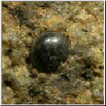
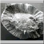
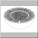
Pachytheca, a peculiar, vegetable little sphereSometimes small glossy spheres are found in sediments of the Upper Silurian and the Lower Devonian. They measure 1 to 6 mm and possess a characteristic internal structure. Hooker named them Pachytheca in 1853, meaning 'thick sporangium'. In the first half of the 19th century they were considered to be seedlike objects or parts of fish jaws. From 1889 on Hooker took the little spheres for algae or colonies of algae and nowadays they are positioned in the Nematophytes, a group of enigmatic organisms, consisting of variously shapes and sized tubes. Other members of this group are Prototaxites, Nematothallus and Nematoplexus.
Pachytheca is most frequently found in the countries of western
Europe: Scotland, Wales, England, Belgium, France and Germany, but there
are also finds from other parts of the world, like Canada and
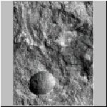
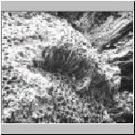
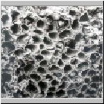
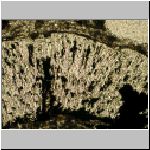
Australia. SEM-photos of Pachytheca from Lac de la Gileppe, Belgium
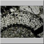
The cortex consists of thin, straight tubes in which sometimes a kind
of thread appears. Tubes occur also in the inner zone, but these are strongly
curved and run in all directions. They are in most cases poorly preserved
indicating a less solid structure. This could be the reason that many specimens
of Pachytheca are found with an empty or sedimentfilled inner
zone.
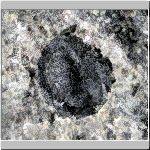
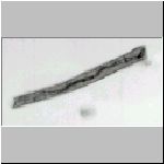
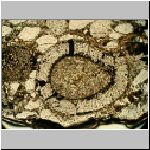
The specimens of Lac de la Gileppe in Belgium have
been so well preserved that they often show the detailed internal structure.
For that purpose a peel can be made from the fossil. A piece of rock
with Pachytheca in it, is sawn through and polished. The polished
surface is then etched with diluted hydrochloric acid. In this way a thin
layer of chalk is removed whereas the organic parts remain. After rinsing
and drying aceton is poored over the etched surface and a piece of cellulose
acetate folium is put over it. This is pressed to the surface and pulled
off after some drying time. The organic remains are now on the peel and they
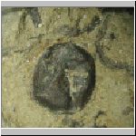
can be studied under the microscope. Photos of peels of Pachytheca from Lac de la Gileppe
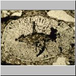
It looks as if the cortex grew relatively thinner as the organism became
older (and more mature?). In young specimens the cortex is about as thick
as the inner zone; in older and bigger specimens the cortex is relatively
much thinner. Pachytheca belongs to a group of enigmatic plants, which conquered the land at about the same time as higher plants (like Cooksonia) did. Other members of this group are Nematothallus, Parka, Prototaxites and Nematoplexus. All plants of these group became extinct during the Devonian. |